Breast Anatomy Quadrants / female breast anatomy and physiology - They rest on the major chest muscle, the pectoralis major.
Get link
Facebook
Twitter
Pinterest
Email
Other Apps
Breast Anatomy Quadrants / female breast anatomy and physiology - They rest on the major chest muscle, the pectoralis major.. Breast quadrants for the anatomical location and description of tumors and cysts, the surface of the breast is divided into four quadrants (fig. • upper outer (superolateral) quadrant • upper inner (superomedial. Lymphatics from the left breast ultimately terminate in the thoracic duct and the left subclavian vein, and from the right breast in the right subclavian vein. 2:00 in the right breast is in the uiq, whereas 2:00 in the left breast is in the uoq. Fat accounts for its smooth contour and most of its bulk.
Lateral quadrants of the breast; Code the primary site to c509 when there are multiple tumors (two or more) in at least two quadrants of the breast. The breast is somewhat circular in shape. For the anatomical location and description of tumors, the surface of the breast is divided into 4 quadrants, upper outer, lower outer, upper inner and lower inner. • upper outer (superolateral) quadrant • upper inner (superomedial.
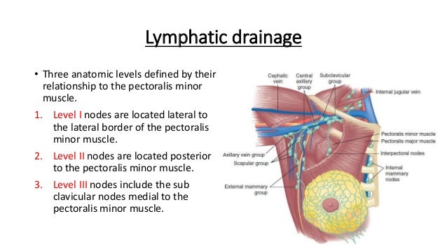
Breast Anatomy from image.slidesharecdn.com Mainly to the axillary lymph nodes as mentioned above. Each of these 4 regions is called a quadrant. They are supported by and attached to the front of the chest wall on either side of the breast bone or sternum by ligaments. • upper outer (superolateral) quadrant • upper inner (superomedial. Another way to describe a breast location is by using the clock face method, in which the location of breast findings is described as though a clock were superimposed on each breast as. This illustration shows the makeup of breast anatomy both inside and outside. Anatomy of the breast for medical/dental students. These lobules are separated by fibrous septa running from the subcutaneous tissues to the fascia of the chest wall.
Code the primary site to c509 when there are multiple tumors (two or more) in at least two quadrants of the breast.
The breast has no muscle tissue.a layer of fat surrounds the glands and extends throughout the breast. These guidelines pertain to the data item grade. Lateral quadrants of the breast; Learn about breast anatomy so you can better understand breast cancer, be aware of anything unusual, & have better dialogue with your doctor. Cage thoracique et sein (illustrations). All genders can get breast cancer. 5 groups of axillary lymph nodes which drain the breast Dominant pathway (receives >75% of lymph from breasts) drains lateral quadrants of breast either directly or via sappey's plexus to axillary nodes An understanding of the basic anatomy, physiology, and histology is important in the. Anatomy of the breast for medical/dental students. Situation and extend • breast is divided in four quadrants 1. Positions of findings in the breast are described in breast quadrants, with the upper outer quadrant representing the breast quadrant nearest the axilla. They rest on the major chest muscle, the pectoralis major.
For the anatomical location and description of tumors, the surface of the breast is divided into 4 quadrants, upper outer, lower outer, upper inner and lower inner. These guidelines pertain to the data item grade. Lymphatics from the left breast ultimately terminate in the thoracic duct and the left subclavian vein, and from the right breast in the right subclavian vein. Positions of findings in the breast are described in breast quadrants, with the upper outer quadrant representing the breast quadrant nearest the axilla. The breast anatomy of males and females is slightly different.
Breast Quadrants Medical Illustration Medivisuals from medivisuals1.com Breast primary with positive nodes and no breast mass found: Cage thoracique et sein (illustrations). Breast shape breast shape and size depend upon genetic, racial and dietary factors, and the age, parity and the main bulk of the breast tissue is usually localized to its upper outer quadrant. In addition, there are also suspensory cooper's ligaments and connective tissue such as collagen and elastin. This illustration shows the makeup of breast anatomy both inside and outside. Theory holds that the upper outer quadrant of the breast develops more malignancies because of increased tissue volume. These guidelines pertain to the data item grade. The breast anatomy of males and females is slightly different.
Lymphatics from the left breast ultimately terminate in the thoracic duct and the left subclavian vein, and from the right breast in the right subclavian vein.
An understanding of the basic anatomy, physiology, and histology is important in the. For the anatomical location and description of tumors, the surface of the breast is divided into 4 quadrants, upper outer, lower outer, upper inner and lower inner. Learn about breast anatomy so you can better understand breast cancer, be aware of anything unusual, & have better dialogue with your doctor. They rest on the major chest muscle, the pectoralis major. The breast is divided into four quadrants: Laterality laterality must be coded for all subsites. The breast has no muscle tissue.a layer of fat surrounds the glands and extends throughout the breast. The breast has an inhomogeneous structure which is predominantly composed of adipose tissue and glandular tissue. In addition, there are also suspensory cooper's ligaments and connective tissue such as collagen and elastin. Lymphatics from the left breast ultimately terminate in the thoracic duct and the left subclavian vein, and from the right breast in the right subclavian vein. This illustration shows the makeup of breast anatomy both inside and outside. Cancer registration & surveillance modules. Another way to describe a breast location is by using the clock face method, in which the location of breast findings is described as though a clock were superimposed on each breast as.
Lymphatics from the left breast ultimately terminate in the thoracic duct and the left subclavian vein, and from the right breast in the right subclavian vein. All genders can get breast cancer. The breast is divided into four quadrants: The breast has an inhomogeneous structure which is predominantly composed of adipose tissue and glandular tissue. Situation and extend • breast is divided in four quadrants 1.
Functional anatomy of cancer from image.slidesharecdn.com Learn about breast anatomy so you can better understand breast cancer, be aware of anything unusual, & have better dialogue with your doctor. This is because this area has a lot of glandular tissue. Lateral quadrants of the breast; Suspended from anterior chest by ligaments of cooper (wikipedia glandular tissue is most abundant in upper outer quadrant of breast; The breast is somewhat circular in shape. In addition, there are also suspensory cooper's ligaments and connective tissue such as collagen and elastin. These guidelines pertain to the data item grade. The four quadrants are upper lateral, upper medial, lower medial, and lower lateral quadrants.
In addition, there are also suspensory cooper's ligaments and connective tissue such as collagen and elastin.
This illustration shows the makeup of breast anatomy both inside and outside. Anatomy of the breast 1. Lateral quadrants of the breast; Mainly to the axillary lymph nodes as mentioned above. Fat accounts for its smooth contour and most of its bulk. They are present in both males and females, yet are more prominent in females following puberty. • upper outer (superolateral) quadrant • upper inner (superomedial. Positions of findings in the breast are described in breast quadrants, with the upper outer quadrant representing the breast quadrant nearest the axilla. The results of this anatomical study may facilitate sn biopsy in patients with breast cancer. In seven cases (29%), the sn was located in the upper ventral quadrant, in two cases (8%) in the upper dorsal quadrant, and in one case in the lower dorsal quadrant. These lobules are separated by fibrous septa running from the subcutaneous tissues to the fascia of the chest wall. The breast has an inhomogeneous structure which is predominantly composed of adipose tissue and glandular tissue. Code the primary site to c509 when there are multiple tumors (two or more) in at least two quadrants of the breast.
These lobules are separated by fibrous septa running from the subcutaneous tissues to the fascia of the chest wall anatomy quadrants. Mainly to the axillary lymph nodes as mentioned above.
Spdf / Advanced Chemistry Atomic Structure Spdf Electronic Configurations Youtube . The space physics data facility (spdf) hosts the s3c active archive, which consists of web services for survey and high resolution data, trajectories, and scientific models. By spdf configuration, he meant orbital configuration. The form of the periodic table is closely related to the electron configuration of the atoms of the elements. For example, all the elements of group 2 have an electron configuration of e ns 2 (where e is an inert gas configuration), and have notable similarities in their chemical properties. To reach spdf support services staff please use our new email address: Now the basic of this concept is from very fundamental quantum chemistry formulation but i don't think you need to know that now (it is usually taught in 4th year of ug or 1st year of graduate study in engineering discipline but i am not sure about pure science discipline). By spdf configuration, he
Pates Italiennes Bio Haut De Gamme : Primavera #Barilla en platillos con Barilla, muchas formas ... - Nous travaillons avec des produits bio et gourmands. . Il y a quelques années. Camargue production cultive et récolte depuis 3 générations les meilleurs riz de camargue. La famille d'appartenance est celle des pâtes courtes et à. Voir plus d'idées sur le thème pates italiennes, recette, cuisine. Les pâtes plaisent autant aux petits qu'aux plus grands. Découvrez les formes, les semoules et les procédés de fabrication. Agidra a sélectionné la gamme bio de la marque italienne 1881 : Mangiobevo propose une large gamme de pâtes, toutes strictement faites à la main par les meilleurs fabricants de pâtes italiennes. Découvrez notre plus belle sélection de pâtes italiennes sèches, fraîches ainsi que du riz pour vos risottos italiens favoris! Cependant, les ingrédients chimiques utilisés dans les produits de beauté traditionnels inquiètent de plus en plus, et de no


Comments
Post a Comment